Home
/ Animal Cell Microscope Experiment / Mitosis Through The Microscope Advances In Seeing Inside Live Dividing Cells Science / Next, look at the prepared cheek side and draw what you see in your notebook.
Animal Cell Microscope Experiment / Mitosis Through The Microscope Advances In Seeing Inside Live Dividing Cells Science / Next, look at the prepared cheek side and draw what you see in your notebook.
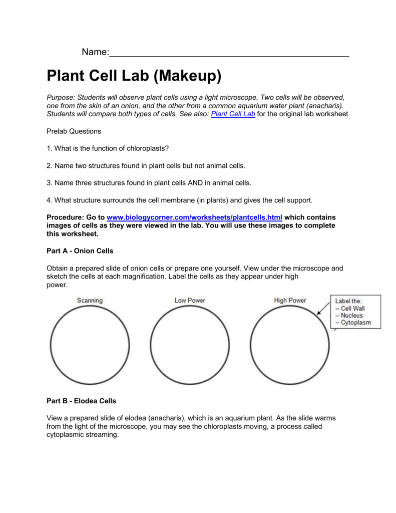
Animal Cell Microscope Experiment / Mitosis Through The Microscope Advances In Seeing Inside Live Dividing Cells Science / Next, look at the prepared cheek side and draw what you see in your notebook.. Rinse your mouth thoroughly before scraping to remove excess food and bacterial deposits.lightly scrape the inner surface of your cheek using the flat side of a toothpick. Onion and cheek cells were observed under optical microscope at a magnification of 100x, 400x and 1000x. The virtual lab begins at the step where you place the slide on the microscope page. For the lesson 9 science experiment from the botany unit from the good an. Comparing plant and animal cells pre lab discussion:
After hundreds of years of observations by many biologists, the cell theory was developed. Once slides have been prepared, they can be examined under a microscope. In order to see these cells they had to be stained with a biological stain. This appears at the light microscope level as a duplication of chromosomes. Cells are taken from the inside cheek of a student.
Plant Cell Virtual Lab from s3.studylib.net In this experiment, stained plant cells After the swabs and slides have been used, they need to be soaked in disinfectant and disposed of according to cleapss recommendations. Microscope is used extensively in cell biology, microbiology, biotechnology, microelectronics, nanophysics, pharmacology, mineralogy, and forensics. One science experiment done, a ton more to go! Animal cells place a small drop of methylene blue on a clean slide. Purchase from tpt science interactive journal unit 1: Two slides demonstrating the cell membrane of an animal cell and the cell wall of a plant cell. Compare animal cells with plant cells e.
The cell theory states that the cell is the structural and.
Swirl the end of the toothpick with the scrapings in the methylene blue on the slide. Four basic types of tissues are found in animals. This subject is important because in biology, we will be using the microscope many times during different laboratory exercises. One of the first scientists to look at cells under a microscope was an english scientist by the name of robert hooke. In this experiment, optical microscope was used to observe the onion skin cells and human cheek cells. Investigating cells with a light microscope. I've had quite a bit of success with. In the late 17th century an englishman, robert hooke, discovered the honeycomb structure or 'cells of a cork when viewing them under his microscope. During this experiment, you will work as a team to use the proscope digital usb microscope and a computer to collect microscopic images from a variety of. First, to prepare an animal cell slide, start by pouring 30 ml of distilled water into a beaker. Comparing plant and animal cells pre lab discussion: One science experiment done, a ton more to go! Two slides demonstrating the cell membrane of an animal cell and the cell wall of a plant cell.
Students will discover that their skin is made up of cells. 6.0 conclusion preparation of 'wet mount' for plant cell, allium sp. After hundreds of years of observations by many biologists, the cell theory was developed. For the lesson 9 science experiment from the botany unit from the good an. In this simple microscope experiment, we will compare plant cells and animal cells.
Plant Cell Virtual Lab from s3.studylib.net Two slides demonstrating the cell membrane of an animal cell and the cell wall of a plant cell. Students will observe cheek cells under a microscope. In the late 17th century an englishman, robert hooke, discovered the honeycomb structure or 'cells of a cork when viewing them under his microscope. In this experiment, stained plant cells We also want to observe the cell shape and several organelles of the cells. Comparing plant and animal cells in this investigation you will use a virtual microscope to view slides of cork cells, onion bulb epidermis cells, privet leaf cells and cheek cells. The animal cells being used in this experiment were squamous epithelial cells obtained from the inside of the human cheek. Microscopy and the study of tissues.
During mitosis, the two sets of chromosomes are precisely separated and each daughter cell receives one complete set.
The animal cells being used in this experiment were squamous epithelial cells obtained from the inside of the human cheek. Comparing plant and animal cells in this investigation you will use a virtual microscope to view slides of cork cells, onion bulb epidermis cells, privet leaf cells and cheek cells. As for the animal cell, instead of extracting the sample from the tongue, the sample might be extracted from the cheeks, with a twist of change of the materials used, where the sample might be extracted by wiping a cotton bud along the inner corners of the cheeks which prevents any harm of the tongue and the vision under the microscope would be. The cell theory states that the cell is the structural and. Onion and cheek cells were observed under optical microscope at a magnification of 100x, 400x and 1000x. The blue helps you see the cells which are normally a clear color. Students will discover that onions are made up of cells. To use a light microscope to examine animal or plant cells Carolina biological supply has everything you need to complete your classroom environmental science experiments. One science experiment done, a ton more to go! This practical involves looking at animal (human) cells through a microscope. After the swabs and slides have been used, they need to be soaked in disinfectant and disposed of according to cleapss recommendations. Purchase from tpt science interactive journal unit 1:
Cheek cells are eukaryotic cells (cells that contain a nucleus and other organelles within enclosed in a membrane) that are easily shed from the mouth lining. Once slides have been prepared, they can be examined under a microscope. Purchase from tpt science interactive journal unit 1: During this experiment, you will work as a team to use the proscope digital usb microscope and a computer to collect microscopic images from a variety of. It's therefore easy to obtain them for observation.
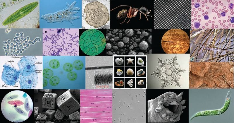
How These 26 Things Look Like Under The Microscope With Diagrams from microbenotes.com Once slides have been prepared, they can be examined under a microscope. Introduction the purpose of this lab was to use the microscope and identify cells such as animal cells and plant cells. Investigating cells with a light microscope. 6.0 conclusion preparation of 'wet mount' for plant cell, allium sp. The null hypothesis of the experiment is that there will be no difference in structure between the two cell types. After the swabs and slides have been used, they need to be soaked in disinfectant and disposed of according to cleapss recommendations. Observing plant and animal cells. After hundreds of years of observations by many biologists, the cell theory was developed.
The compound light microscope that is used in this lab allows you to see small, eukaryotic organisms or thinly sliced sections of
It was hooke who coined the term 'cells'. Finally, that the individual cells will be approximately 0.05mm in length/diameter. Next, look at the prepared cheek side and draw what you see in your notebook. The blue helps you see the cells which are normally a clear color. Students will observe onion cells under a microscope. Comparing plant and animal cells pre lab discussion: Plant and animal cells microscope lab. During this experiment, you will work as a team to use the proscope digital usb microscope and a computer to collect microscopic images from a variety of. Using the microscope & cell biology the light microscope is able to magnify and then focus visible light energy by using lenses. During mitosis, the two sets of chromosomes are precisely separated and each daughter cell receives one complete set. We also want to observe the cell shape and several organelles of the cells. First, to prepare an animal cell slide, start by pouring 30 ml of distilled water into a beaker. The null hypothesis of the experiment is that there will be no difference in structure between the two cell types.
Share :
Post a Comment
for "Animal Cell Microscope Experiment / Mitosis Through The Microscope Advances In Seeing Inside Live Dividing Cells Science / Next, look at the prepared cheek side and draw what you see in your notebook."

Post a Comment for "Animal Cell Microscope Experiment / Mitosis Through The Microscope Advances In Seeing Inside Live Dividing Cells Science / Next, look at the prepared cheek side and draw what you see in your notebook."