Home
/ Diagram Of An Animal Cell Under An Electron Microscope - Rana Ray Diagram Of Animal Cell Seen Through Electron Microscope Brainly In - Animal and plant cell under electron microscope.
Diagram Of An Animal Cell Under An Electron Microscope - Rana Ray Diagram Of Animal Cell Seen Through Electron Microscope Brainly In - Animal and plant cell under electron microscope.
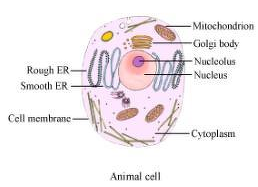
Diagram Of An Animal Cell Under An Electron Microscope - Rana Ray Diagram Of Animal Cell Seen Through Electron Microscope Brainly In - Animal and plant cell under electron microscope.. Here is the microscopic view of animal cell. Respiration:mitochondria protein synthesis:endoplasmic reticulum transport of material :endoplasmic reticulum and golgi bodies. (ii) presence of large central vacuole in plant cell. The animal cell is more. But at the same time it is interpretive.
Click (or tap) the diagram for a simple labelled version. Electron microscope constructed by ernst ruska in 1933. In addition the optical and electron microscope, scientists are able to use a number of other techniques to probe. Draw a neat diagram of plant cell and label any three parts which differentiate it from animal cell. How is it different from animal cell?
Lysosomes Dr Jastrow S Electron Microscopic Atlas from www.drjastrow.de How is it different from animal cell? Slides and how to plant and animal cells can be studied in greater detail with a light microscope by magnifying the image. However, when you use an electron microscope to increase the magnification many thousands of times you see that these seemingly simple structures are incredibly complex, each with its own specialized function. Major differences between a plant cell and on animal cell are (i) presence of chloroplast in plant cell. Given below is the diagram of a cell as seen under the microscope after having been placed in a solution Electron microscope constructed by ernst ruska in 1933. After this, add another oval shape outside the line you just drew, and this will make the cell membrane to your animal cell. An animal cell as seen with an electron microscope.
Electron microscope constructed by ernst ruska in 1933.
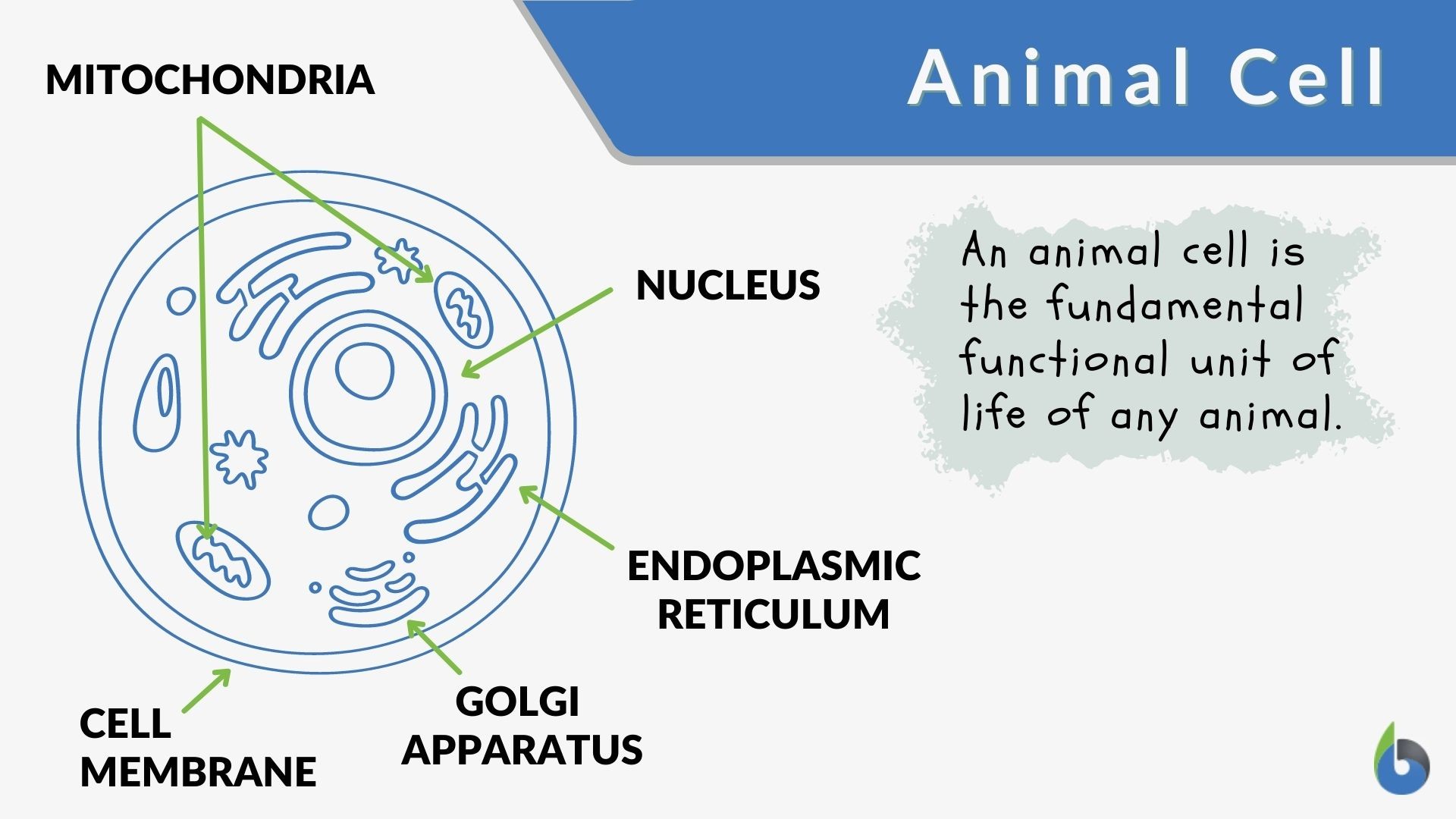
Plant and animal cells have a nucleus inside the cytoplasm. Ishita observed a slide of eukaryotic cell under electron microscope. Given below is the diagram of a cell as seen under the microscope after having been placed in a solution Some disadvantage of electron microscopes are that they cannot display living specimens in natural colours. Here's a photo of a plant cell under an electron microscope. What does an animal cell look like under an electron. Typical animal cell pinocytotic vesicle lysosome golgi vesicles golgi vesicles rough er (endoplasmic reticulum) smooth er (no ribosomes) cell (plasma) 2. Respiration:mitochondria protein synthesis:endoplasmic reticulum transport of material :endoplasmic reticulum and golgi bodies. Plant, animal and bacterial cells have smaller components each the magnification of a microscope is not the only factor that is important when viewing cells. Difference between animal and plant cell. A.robert hooke:studied cork section and name the. Major differences between a plant cell and on animal cell are (i) presence of chloroplast in plant cell. In addition the optical and electron microscope, scientists are able to use a number of other techniques to probe.
Look at the diagram which identifies the different components in a simple animal cell. The final image produced from an electron when you look at animal or plant cells under the electron microscope, you can see a lot more the diagram shows a phospholipid bilayer (cell membrane) with carbon dioxide molecules on one side of. Unfortunately, wikianswers does notallow for drawing tools; How is it different from animal cell? Studying the structures of a live paramecium 4.
Q14 Draw A Large Diagram Of An Animal Cell As Seen Through An Electron Microscope Label The Parts That Science Tissues 11500353 Meritnation Com from s3mn.mnimgs.com Cell structure teaching resources the science teacher, organelles biology for majors i, 11 different types of cells in the human body, class test, chronic inflammation under the microscope learn share. However, when you use an electron microscope to increase the magnification many thousands of times you see that these seemingly simple structures are incredibly complex, each with its own specialized function. Cell scanning electron microscope hd stock video 717 725 243. It controls all the processes and chemical reactions that take. Plant, animal and bacterial cells have smaller components each the magnification of a microscope is not the only factor that is important when viewing cells. Plant cells have cell walls, one large vacuole per cell, and chloroplasts, while animal cells will have a cell membrane only. A cell is a very tiny structure which exists in living bodies. Animal and plant cell under electron microscope.
Here's a diagram of a plant cell:
Difference between animal and plant cell. Slides and how to plant and animal cells can be studied in greater detail with a light microscope by magnifying the image. Detail study of animal cell under electron microscope. A scale bar has been marked on the. The detail that can be seen, or resolution, is also important. However, when you use an electron microscope to increase the magnification many thousands of times you see that these seemingly simple structures are incredibly complex, each with its own specialized function. Now the first thing to point out when looking at images under an electron microscope is the scale. Here is the microscopic view of animal cell. (ii) presence of large central vacuole in plant cell. Animal cell (as seen under electron microscope). The animal cell is more. The diagram is very clear, and labeled the diagram is very clear, and labeled; Ppt structure of plant and animal cells under an electron.
We say cells are microscopic because they can only be seen under a microscope. Which cell structure can only be seen with an electron microscope aice? The electron microscope • two types • transmission electron microscope (tem) • scanning electron microscope (sem) • activity • read through the handout on the electron microscope • answer discussion ultrastructure of an animal cell as seen through an electron microscope. Typical animal cell pinocytotic vesicle lysosome golgi vesicles golgi vesicles rough er (endoplasmic reticulum) smooth er (no ribosomes) cell (plasma) 2. Electron micrographs are sometimes shown in colour.
Animal Cell Definition And Examples Biology Online Dictionary from www.biologyonline.com Electron microscope constructed by ernst ruska in 1933. Cell structure teaching resources the science teacher, organelles biology for majors i, 11 different types of cells in the human body, class test, chronic inflammation under the microscope learn share. After this, add another oval shape outside the line you just drew, and this will make the cell membrane to your animal cell. In addition the optical and electron microscope, scientists are able to use a number of other techniques to probe. Draw a neat diagram of plant cell and label any three parts which differentiate it from animal cell. Plant, animal and bacterial cells have smaller components each the magnification of a microscope is not the only factor that is important when viewing cells. Given below is the diagram of a cell as seen under the microscope after having been placed in a solution Cell membrane dr jastrow s electron microscopic what does an animal cell look like under an electron microscope.
In addition the optical and electron microscope, scientists are able to use a number of other techniques to probe.
Plant, animal and bacterial cells have smaller components each the magnification of a microscope is not the only factor that is important when viewing cells. The parts that carry out the functions are: The diagram is very clear, and labeled the diagram is very clear, and labeled; The electron microscope two main advantages high resolving power (short wavelength of electrons) as electrons negatively are charged the beam can be focused using electromagnets as electrons are absorbed by molecules 7 ultrastructure of an animal cell as seen through an electron microscope. Which cell structure can only be seen with an electron microscope aice? Electron microscopes use accelerated electron beams (as opposed to visible light in a light microscope) to create images of magnification as here is an electron micrograph of an animal cell with the labels superimposed: Respiration:mitochondria protein synthesis:endoplasmic reticulum transport of material :endoplasmic reticulum and golgi bodies. Given below is the diagram of a cell as seen under the microscope after having been placed in a solution Light and electron microscopes allow us to see inside cells. However, when you use an electron microscope to increase the magnification many thousands of times you see that these seemingly simple structures are incredibly complex, each with its own specialized function. Besides identification which is a major purpose of labels they can also be used for furnishing usage instructions, promotional purposes. Unfortunately, wikianswers does notallow for drawing tools; Cell structure teaching resources the science teacher, organelles biology for majors i, 11 different types of cells in the human body, class test, chronic inflammation under the microscope learn share.
But at the same time it is interpretive diagram of an animal cell under electron microscope. Electron microscopes use electron beams focused by electromagnets to magnify and resolve microscopic specimens.
Share :
Post a Comment
for "Diagram Of An Animal Cell Under An Electron Microscope - Rana Ray Diagram Of Animal Cell Seen Through Electron Microscope Brainly In - Animal and plant cell under electron microscope."


Post a Comment for "Diagram Of An Animal Cell Under An Electron Microscope - Rana Ray Diagram Of Animal Cell Seen Through Electron Microscope Brainly In - Animal and plant cell under electron microscope."