Home
/ Electron Microscope Diagram Of Animal Cell / Structure of Animal Cell and Plant Cell Under Microscope ... : Given below is the diagram of a cell as seen under the microscope after having been placed in a solution
Electron Microscope Diagram Of Animal Cell / Structure of Animal Cell and Plant Cell Under Microscope ... : Given below is the diagram of a cell as seen under the microscope after having been placed in a solution
Electron Microscope Diagram Of Animal Cell / Structure of Animal Cell and Plant Cell Under Microscope ... : Given below is the diagram of a cell as seen under the microscope after having been placed in a solution. Electron microscopes use electron beams focused by electromagnets to magnify and resolve microscopic specimens. (b) diagram of the clairscope. However, when you use an electron microscope to increase the magnification many thousands of times you see that these seemingly simple structures are incredibly complex, each with its own specialized function. (iii) presence of cell wall. A.robert hooke:studied cork section and name the.
Electron microscopy is routinely used as a tool in such diverse areas as anatomy, anthropology the lenses in electron microscopes are windings of copper wire, or solenoids, which generate a secondary light electron guide collector. The ultrastructure of cells viewed by transmission electron microscopy and scanning electron microscopy. These diagrams clearly explain the functioning of the microscopes along with their respective parts. Cells are the basic units of structure and function in living things. The type of cell and the structure of cells.
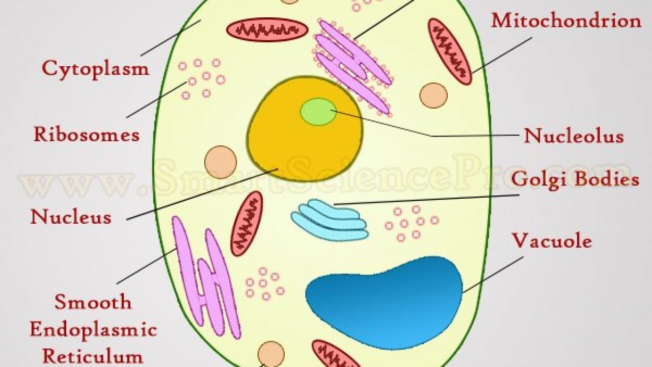
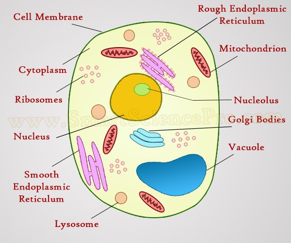
Labelled Diagram Of A Plant Cell Under A Microscope ... from www.smartsciencepro.com The type of cell and the structure of cells. Difference between animal and plant cell. Click (or tap) the diagram for a simple labelled version. In a transmission electron microscope, the electron beam penetrates the cell and provides plant cells have a cell wall, chloroplasts, plastids, and a central vacuole—structures not found in animal cells. As the wavelength of an electron can be up to 100. A cell surface membrane b chromosome c nucleolus d vacuole. Cells are the basic units of structure and function in living things. Look at the diagram which identifies the different components in a simple animal cell.
This is because of the way that the cell was sectioned (cut) before it was viewed on the transmission electron microscope.
Light and electron microscopes allow us to see inside cells. Given below is the diagram of a cell as seen under the microscope after having been placed in a solution Here is an electron micrograph of an animal cell with the labels superimposed: Click (or tap) the diagram for a simple labelled version. When you look at animal or plant cells under the electron microscope, you can see a lot more detail. Cells are the basic units of structure and function in living things. The type of cell and the structure of cells. An electron microscope is a microscope that uses a beam of accelerated electrons as a source of illumination. Figure 1 diagram showing the basic components making. The diagram shows several processes taking place in a cell. These diagrams clearly explain the functioning of the microscopes along with their respective parts. However, when you use an electron microscope to increase the magnification many thousands of times you see that these seemingly simple structures are incredibly complex, each with its own specialized function. But at the same time it is interpretive.
(iii) presence of cell wall. Anatomy_and_physiology_of_animals_animal_cell_electron_microscope.jpg (557 × 540 pixels, file size: It is much stronger than a light microscope so is used to take pictures of extremely small. All cells are produced from other cells. Most of the cells are microscopic hence they can only be seen under a microscope in order to.
A LEVEL SCIENCE NOTES: Plant cell structure from 1.bp.blogspot.com You see that many features are in common. Which cell structure can be seen only with an electron microscope? Electron microscopy is routinely used as a tool in such diverse areas as anatomy, anthropology the lenses in electron microscopes are windings of copper wire, or solenoids, which generate a secondary light electron guide collector. Of an animal cell and its this transmission electron. (ii) presence of large central vacuole in plant cell. Respiration:mitochondria protein synthesis:endoplasmic reticulum transport of material :endoplasmic reticulum and golgi bodies. Ultrastructure is the architecture of cells that is visible at higher magnifications than found on a standard light microscope. It occurs when the electrons interact with the cell nuclei;
The type of cell and the structure of cells.
An animal cell as seen with an electron microscope. Unlike the eukaryotic cells of plants and fungi, animal cells do not have a cell wall. What invention made it possibe for people uses a beam of electrons to produce a magnified image; The detail that can be seen, or resolution, is also important. Which cell structure can be seen only with an electron microscope? However, when you use an electron microscope to increase the magnification many thousands of times you see that these seemingly simple structures are incredibly complex, each with its own specialized function. You see that many features are in common. All cells are produced from other cells. Given below is the diagram of a cell as seen under the microscope after having been placed in a solution Typical animal cell pinocytotic vesicle lysosome golgi vesicles golgi vesicles rough er (endoplasmic reticulum) smooth er (no ribosomes) cell (plasma) 2. It is much stronger than a light microscope so is used to take pictures of extremely small. Click (or tap) the diagram for a simple labelled version. (ii) presence of large central vacuole in plant cell.
The electron microscope is a compound microscope in which the arrangement of the main lenses follows the same pattern as in the light microscope fig. Animal cell definition with cell size and shape. Here is an electron micrograph of an animal cell with the labels superimposed: When viewed with an electron microscope, the cylinders show up as nine bundles of tiny. Given below is the diagram of a cell as seen under the microscope after having been placed in a solution
Structure of Animal Cell and Plant Cell Under Microscope ... from www.smartsciencepro.com It occurs when the electrons interact with the cell nuclei; You see that many features are in common. Cells are the basic units of structure and function in living things. Of an animal cell and its this transmission electron. The ultrastructure of cells viewed by transmission electron microscopy and scanning electron microscopy. An animal cell as seen with an electron microscope. We say cells are microscopic because they can only be seen under a microscope. The type of cell and the structure of cells.
Here is the microscopic view of animal cell.
This is because of the way that the cell was sectioned (cut) before it was viewed on the transmission electron microscope. As the wavelength of an electron can be up to 100. The diagram shows several processes taking place in a cell. You see that many features are in common. A ciliated epithelial cell b goblet cell c red blood cell d smooth muscle cell. Unlike the eukaryotic cells of plants and fungi, animal cells do not have a cell wall. Figure 1 diagram showing the basic components making. Figure 7.14 at left a transmission electron micrograph and at right a labeled diagram of a. An electron microscope is a microscope that uses a beam of accelerated electrons as a source of illumination. Both light microscopes and electron microscopes use radiation (light or electron beams) to form the following simple block diagram shows some of the basic similarities between light microscopes and electron microscopes (in general) by comparing. Light microscope vs electron microscope. Respiration:mitochondria protein synthesis:endoplasmic reticulum transport of material :endoplasmic reticulum and golgi bodies. When you look at animal or plant cells under the electron microscope, you can see a lot more detail.
Share :
Post a Comment
for "Electron Microscope Diagram Of Animal Cell / Structure of Animal Cell and Plant Cell Under Microscope ... : Given below is the diagram of a cell as seen under the microscope after having been placed in a solution"


Post a Comment for "Electron Microscope Diagram Of Animal Cell / Structure of Animal Cell and Plant Cell Under Microscope ... : Given below is the diagram of a cell as seen under the microscope after having been placed in a solution"